
ORIGINAL ARTICLE
TECHNICAL PROTOCOL FOR WOUND PHOTOGRAPHIC DOCUMENTATION: A TECHNOLOGICAL DEVELOPMENT STUDY*
Perla Oliveira Soares de Souza1, Alexandre Cruz Leão2, Josimare Aparecida Otoni Spira3, Eline Lima Borges4
1 Federal University of Minas Gerais, School of Nursing. Belo Horizonte, MG, Brazil. ORCID: 0000-0002-6380-233X. E-mail: ossouzaperla@gmail.com.
2 Federal University of Minas Gerais, School of Fine Arts. Belo Horizonte, MG, Brazil. ORCID: 0000-0003-1902-2165. E-mail: alexandre.leao.ufmg@gmail.com.
3 Federal University of Minas Gerais, School of Nursing. Belo Horizonte, MG, Brazil. ORCID: 0000-0002-6577-0394. E-mail: j.otoni@yahoo.com.br.
4 Federal University of Minas Gerais, School of Nursing. Belo Horizonte, MG, Brazil. ORCID: 0000-0002-0623-5308. E-mail: elineufmg@gmail.com.
ABSTRACT
Objective: To develop a technical protocol for standardizing photographic wound documentation in the clinical care context. Method: A technological development study conducted according to the SQUIRE 2.0 principles. Initially, a structured literature search was performed to establish technical parameters related to clinical wound photography. Based on the identified evidence, a preliminary version of the protocol was drafted. Subsequently, the instrument underwent technical evaluation in a controlled environment at the Laboratory of Scientific Documentation by Image of a Brazilian public university. Aspects such as camera positioning, distance, lighting, framing, use of a metric scale, color calibration, and image storage were analyzed, focusing on operational feasibility and technical reproducibility. Results: The evaluation allowed for the simplification of recommendations and adjustments to ensure practical applicability in the healthcare setting. The final protocol was organized into a checklist format, structured across three dimensions: preparation for documentation, the photographic act, and image storage and security, aiming for standardization, documentary traceability, and technical consistency. Conclusion: The developed protocol constitutes a structured tool for qualifying wound photographic documentation, contributing to the reduction of technical variability and the strengthening of care quality and clinical governance.
Descriptors: Wounds and Injuries; Photography; Documentation; Nursing; Quality Improvement; Clinical Governance.
|
How to cite: Souza POS, Leão AC, Spira JAO, Borges EL. Technical protocol for wound photographic documentation: a technological development study. Online Braz J Nurs. 2026;25(1):e20266960. http://doi.org/10.17665/1676-4285.20266960 |
What is already known:
Photographic documentation is widely used in wound monitoring, but it presents technical variability related to lighting, angulation, and framing.
The lack of standardization compromises the serial comparability of the images and may affect the quality of clinical documentation.
There is a gap in structured technical protocols aimed at nursing care practice in the Brazilian context.
What this article adds:
The development of a structured technical protocol for standardizing wound photographic documentation, organized as an operational checklist.
The integration of technical, ethical, and organizational aspects, contributing to the improvement of care quality and health information governance.
The provision of an applicable instrument to reduce technical variability and strengthen documentary traceability in clinical practice.
INTRODUCTION
The systematic assessment of chronic wounds is an essential component of healthcare, particularly in nursing. The nurse plays a central role in the longitudinal follow-up of these patients(1). Clinical monitoring involves the analysis of dimensions, tissues present, exudate, borders, perilesional skin, and signs of infection, which support therapeutic decisions and reassessments of the care plan.
The use of standardized photographic documentation has become consolidated as a complementary tool to written documentation, allowing for serial comparative analysis, communication among professionals, support for teleassistance, and ethical-legal backing throughout the care process. Recent clinical studies and reviews emphasize the role of digital imaging as a means to capture changes in wound morphology and to support quantitative and qualitative assessments of healing evolution—especially when conventional methods heavily depend on the operator and exhibit variability in the precision and reliability of measurements(2).
Technological advances have significantly expanded access to image-capture devices, particularly high-resolution smartphones, which represent a promising resource(3). However, technological availability alone does not ensure technical quality or methodological standardization. Evidence indicates frequent inconsistencies in camera positioning, lighting variability, absence of metric scales, and failures in image documentation and storage, thereby compromising clinical reliability and information security(4). This lack of consistent technical protocols for the photographic documentation of chronic wounds undermines both the quality of clinical records and the comparability of serial assessments across different healthcare professionals and services(5).
In the literature, protocol-based care is understood as an approach that organizes and standardizes professional practice through previously defined structures, promoting consistency and quality of assistance processes, in addition to reducing variability in clinical practice (6). Protocols aimed at executing procedures, such as the photographic registration of wounds, assume a technical character by establishing steps and parameters for their execution, contributing to the standardization of practice and the quality of clinical records, especially when based on structured image capture criteria(7). In this sense, the term “technical protocol” is adopted in this study to designate instruments focused on standardizing the execution of procedures through the definition of technical steps and parameters.
Beyond the technical standardization established by the protocol, wound photographic documentation involves ethical and legal dimensions. Obtaining informed consent, protecting sensitive data, and the institutional use of digital devices are emerging themes in the literature, especially in the context of the digitalization of clinical records and the integration of mobile health platforms(8).
Despite the recognized importance of these elements, there are significant gaps in the literature concerning methodologically robust and validated protocols for standardizing wound photographic documentation within the nursing context. Specifically, studies that describe the development and prior testing of technical parameters before implementation in real clinical settings(9).
Given this gap, it becomes necessary to develop a technological product that organizes recommendations based on scientific literature and submit it to technical evaluation in a controlled environment, with the objective of verifying its viability, technical consistency, and applicability in the care context.
Based on this premise, the present study aims to develop a technical protocol for standardizing wound photographic documentation in the care context.
METHOD
Study design
This is a technological development study with technical evaluation in a controlled environment, conducted according to the principles of the Standards for Quality Improvement Reporting Excellence (SQUIRE 2.0), aimed at the systematic description of interventions intended to qualify care processes(10).
The study aimed to develop a technical protocol for standardizing wound photographic documentation in the clinical care context.
Context
The evaluation was carried out at the Laboratory of Scientific Documentation by Image of the Federal University of Minas Gerais (iLab-UFMG). This is an environment specialized in the capture and technical management of digital images.
The choice of this setting was justified by the possibility of rigorous control over the technical variables involved in photographic documentation, such as lighting, camera positioning, image background, and color calibration. The control allows for the evaluation of the reproducibility and consistency of the proposed recommendations.
Technical protocol development
The protocol development occurred in two sequential and complementary stages.
In Stage 1 – Technical-Scientific Basis, a structured search of the scientific literature was conducted with the objective of identifying technical parameters related to wound photographic documentation. This stage was instrumental, aiming to support the initial drafting of the protocol.
International and Latin American databases were consulted, including MEDLINE/PubMed, Embase, Scopus, Web of Science, Cochrane Library, and LILACS, using descriptors related to clinical photography, wound documentation, and image standardization.
The identified studies were analyzed for recommendations regarding: camera positioning; distance and angulation; lighting; image background; use of a metric scale; color calibration; image storage and security. The findings were organized into preliminary technical parameters that constituted the initial version of the protocol.
Stage 2 – Protocol Evaluation in a Controlled Environment, consisted of submitting the preliminary version of the protocol to technical evaluation in a controlled environment, using wound simulators. The following were used: two wound simulators; institutional device for image capture; lighting system with LED sources; standardized background; auxiliary instruments for color calibration.
The evaluation aimed to: analyze the technical performance of the recommendations; test the reproducibility of the images; identify technical inconsistencies; adjust or suppress recommendations unfeasible in the care context.
During this stage, the recommendations were reorganized, adapted, or excluded according to criteria of practical applicability, clarity, and technical consistency. From this process, the technical protocol was consolidated and organized into an operational checklist format, structured across three sequential dimensions: preparation for documentation, the photographic act, and post-capture/storage. The checklist was developed with the objective of systematizing the technical recommendations, organizing them into clear steps.
Variables analyzed
In the controlled environment evaluation, the following technical aspects of the produced images were analyzed: sharpness and focus; lighting uniformity; color fidelity; centering of the area of interest; presence of a metric scale; and consistency across serial records. The analysis was descriptive and comparative between different technical configurations.
Data analysis
The obtained data were analyzed descriptively, considering the technical suitability of the generated images and the applicability of the proposed recommendations. The identified inconsistencies supported the consolidation of the final protocol version.
Ethical considerations
As this was a technological development study conducted in a controlled environment, using wound simulators and data derived from scientific literature, with no direct involvement of human subjects, there was no need for submission to a Research Ethics Committee (REC), in accordance with current regulations.
For illustrative purposes of the protocol's applicability, a clinical image was used, which was previously authorized through a specific Informed Consent Form (ICF) for scientific use, without patient identification.
RESULTS
The technical-scientific foundation stage allowed for the identification of parameters related to the photographic documentation of wounds, initially organized into ten structural axes encompassing ethical, technical, and operational aspects. These parameters were grouped into three operational dimensions, structured sequentially, establishing a logical order of the steps to be followed for the proper performance of the photographic record.
1. Preparation for documentation
2. Execution of the photographic record
3. Image storage and security
The preliminary organization supported the construction of the initial version of the technical protocol (Table 1).
Table 1 – Proposed parameters and recommendations for wound photographic documentation. Belo Horizonte, MG, Brazil, 2022
|
Synthesized technical recommendations |
|
|
1) Informed Consent |
Obtain a specific Informed Consent Form for photographic documentation, providing clarification regarding the intended use of the images. |
|
2) Camera Positioning |
Maintain a perpendicular angle (90°) to the wound and a standardized distance (20–30 cm), ensuring visualization of the involved limb and consistency across serial records. |
|
3) Lighting |
Utilize a uniform white light source, maintaining consistency across records. Control flash usage to prevent shadows and specular reflection. Ideally, use two light sources positioned at 45°. |
|
4) Focus and Framing |
Center the area of interest, ensure sharpness, and utilize optical zoom when necessary. For extensive lesions, capture multiple images while maintaining technical standards. |
|
5) Image Background |
Utilize a neutral and discreet background (preferably blue or white) to provide contrast with skin tones. |
|
6) Identification and Scale |
Include standardized identification and a graduated metric scale positioned near the lesion, without distorting or obstructing the area of interest. |
|
7) Patient Preparation and Positioning |
Perform prior wound cleansing, maintain the patient in an anatomical position, and avoid artifacts that may interfere with visualization. |
|
8) Color Consistency |
Maintain consistent imaging parameters; perform white balance calibration according to the equipment and, when possible, utilize a color reference card. |
|
9) Equipment |
Use standardized institutional equipment, maintaining the same configuration for serial records. Avoid the use of personal devices due to security risks. |
|
10) Storage and Security |
Save images to the institutional system, preserve quality (JPG, PNG, or TIFF), and ensure data protection and traceability. |
Source: prepared by the authors, 2022.
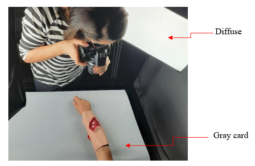
The technical evaluation conducted at the Laboratory of Scientific Documentation by Image (iLab-UFMG) enabled testing the applicability of the recommendations under controlled conditions of lighting, positioning, and capture (Figure 1).
Figure 1 – Controlled environment used for the technical evaluation of the protocol (lighting system, standardized background, and equipment positioning). Belo Horizonte, MG, Brazil, 2022
Source: authors' files, 2026.
During this stage, technical limitations were identified, primarily related to the use of professional photographic equipment, the complexity of color calibration, the use of a gray background, and the need to simplify the operational workflow.
The technical evaluation in a controlled environment allowed for the identification of operational simplification needs and adjustments to ensure the practical feasibility and reproducibility of the preliminary recommendations (Table 2).
Table 2 – Adjustments implemented following the technical evaluation conducted in a controlled environment. Belo Horizonte, MG, Brazil, 2022
|
Parameter |
Preliminary Recommendation |
Implemented Adjustment |
Technical Rationale |
|
Consent |
Provision of Informed Consent Form (ICF) for documentation |
Maintained without changes |
Indispensable ethical-legal requirement |
|
Camera Positioning |
90° angle and 20–30 cm distance |
Reinforced standardization; removal of additional distance variations |
To ensure reproducibility and prevent angular distortions |
|
Lighting |
Use of uniform white light; possibility of flash; two sources at 45° |
Recommendation for uniform white light; use of flash only when necessary; two sources indicated as ideal (not mandatory) |
Operational simplification and feasibility in the care context |
|
Focus and Framing |
Multiple guidelines on zoom and depth |
Synthesis focusing on central focus, sharpness, and preferential use of optical zoom |
Reduction of technical complexity |
|
Identification and Scale |
Extensive detailing on ruler and identification |
Operational synthesis maintaining mandatory scale and standardization |
To facilitate practical application |
|
Color Consistency |
Mandatory use of gray card or color chart |
Made an optional recommendation, according to institutional availability |
Practical feasibility in different settings |
|
Equipment |
Inclusion of professional camera and cell phone |
Exclusion of professional camera; prioritization of standardized institutional equipment |
Technical complexity and low applicability |
|
Storage and Security |
Detailed guidelines on formats and copies |
Synthesis maintaining quality preservation and institutional security |
Focus on traceability and data protection |
Source: prepared by the authors, 2026.
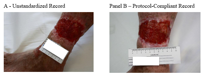
Figure 2 presents an illustrative comparison between the photographic record performed without standardization and that obtained after applying the technical protocol, demonstrating differences related to angulation, framing, the presence of a metric scale, and lighting uniformity.
Figure 2 – Illustrative comparison of wound photographic record before (A) and after (B) application of the technical protocol. Image B shows standardization of perpendicular framing, presence of an adequately positioned metric scale, neutral background without artifacts, and uniform lighting (image is for demonstrative purpose of the protocol's operational applicability). Belo Horizonte, MG, Brazil, 2022
Source: authors' files, 2026.
The consolidated technical protocol was organized into an operational checklist format, structured across three dimensions: preparation for recording, the photographic act, and post-capture/storage (Table 3). This organization aims to facilitate practical applicability in the care context and promote the standardization of serial records.
Table 3 – Technical Protocol for Standardizing Wound Photographic Record – Operational Checklist. Belo Horizonte, MG, Brazil, 2022
|
Item |
Technical Criterion |
Verification (✓) |
|
|
Section 1 – Preparation for Recording |
|
|
1 |
Use standardized institutional equipment |
|
|
2 |
Maintain the same camera configuration for serial records |
|
|
3 |
Ensure uniform white lighting |
|
|
4 |
Use a neutral background (preferably blue or white) |
|
|
5 |
Obtain specific Informed Consent Form (ICF) for photographic recording |
|
|
6 |
Perform prior wound cleansing |
|
|
|
Section 2 – Photographic Act |
|
|
7 |
Position the camera perpendicularly (90°) to the wound |
|
|
8 |
Maintain an approximate distance of 20–30 cm |
|
|
9 |
Center the area of interest |
|
|
10 |
Include the metric scale graduated in centimeters |
|
|
11 |
Position the ruler without pressing the skin |
|
|
12 |
Maintain the patient in an anatomical position |
|
|
13 |
Avoid artifacts (adornments, colored gloves, or shadows) |
|
|
14 |
Ensure focus and sharpness of the area of interest |
|
|
|
Section 3 – Post-Capture and Storage |
|
|
15 |
Save the image in the institutional system |
|
|
16 |
Maintain standardized image identification |
|
|
17 |
Use a format that preserves quality (JPG, PNG, or TIFF) |
|
|
18 |
Ensure data security and protection |
|
|
19 |
Avoid the use of personal devices |
|
|
20 |
Maintain technical consistency in subsequent records |
|
Source: prepared by the authors, 2026.
The checklist was structured to allow sequential application, promoting internal auditing and institutional quality control.
DISCUSSION
The standardization of wound photographic documentation, as proposed in this study, should be understood not merely as a technical refinement but as a structural strategy for quality of care and clinical governance. Variability in the capture of clinical images has been widely documented in international literature, showing inconsistencies related to angulation, lighting, framing, and the absence of a metric scale, factors that compromise the serial comparability and reliability of evolutionary assessments(1,11).
The lack of standardization in photographic documentation can lead to divergent interpretations among professionals, hindering clinical decision-making and the continuity of care. Recent studies on digital wound monitoring demonstrate that the technical quality of the image can influence the precision of tissue assessment and the measurement of the injured area(3,12). Thus, standardization is not limited to aesthetics or documentary organization but constitutes a structural element for reducing inter-observer variability and strengthening clinical consistency.
From the perspective of clinical governance, standardized documentation protocols favor traceability, internal audit, and institutional transparency. The incorporation of operational guidelines for photographic documentation helps to consolidate safe practices for the storage and protection of sensitive data, especially in a scenario of increasing digitalization of electronic health records and the use of mobile devices in care. The literature on digital health emphasizes that clinical information governance requires not only technological infrastructure but also clear standardization of data capture, storage, and sharing processes(13).
In the Brazilian context, COFEN Resolution No. 554/2017 establishes guiding criteria for the use of images by nursing professionals, emphasizing ethical responsibility and patient identity protection(14). However, professional regulation does not detail technical parameters for image capture, a gap that reinforces the relevance of the protocol developed in this study. By integrating technical, ethical, and organizational aspects, the protocol expands the discussion of clinical photography beyond the technological domain, inserting it into the field of patient safety and quality of care.
Another relevant aspect relates to operational feasibility. The exclusion of professional photographic equipment and the simplification of chromatic calibration recommendations reflect the need for adaptation to the care reality. Overly complex protocols tend to have low adherence and limited practical implementation. The literature on health process improvement indicates that sustainable interventions must balance technical rigor and contextual applicability(10). In this sense, the technical evaluation in a controlled environment allowed for adjustments that favor feasibility without compromising methodological consistency.
The organization of the protocol in an operational checklist format also aligns with contemporary strategies for quality improvement, where structured instruments favor standardization, monitoring, and permanent education. Checklists have been widely used in patient safety to reduce variability and promote the reliability of clinical processes(15-16). By adapting this logic to wound photographic documentation, the present study contributes to integrating clinical documentation and quality management. The proposed protocol does not replace the professional's clinical judgment but offers an organizational structure to qualify and standardize care documentation.
Although developed and tested in a controlled environment, the protocol demonstrates potential for implementation in real clinical practice settings. However, future studies should evaluate its applicability in different care contexts, as well as analyze indicators of adherence and impact on documentation quality.
The main limitation of this study refers to the technical evaluation being conducted in a controlled environment, using wound simulators. Although this strategy allowed for rigorous control of variables related to lighting, positioning, and framing, it does not encompass the complexity of real care scenarios, where environmental factors, structural limitations, and anatomical variability may interfere with the protocol's applicability.
Furthermore, the study did not evaluate indicators of professional adherence or measure the impact on clinical outcomes or documentation quality in a care setting. The protocol was developed and adjusted from a technical perspective, and future investigation into its implementation in distinct clinical contexts and its incorporation into institutional systems is necessary.
The developed protocol offers a structured instrument for the standardization of wound photographic documentation, integrating technical, ethical, and organizational aspects. Its organization in a checklist format favors sequential application, internal auditing, and permanent education for the teams.
By proposing clear technical parameters for the capture and storage of clinical images, the study contributes to strengthening the quality of care, documentary traceability, and health information governance. The standardization of photographic documentation can support accreditation processes, patient safety, and continuity of care, especially in contexts of increasing digitalization of electronic health records.
CONCLUSION
The study enabled the development of a technical protocol for standardizing wound photographic documentation, structured based on scientific recommendations and adjusted to present feasibility in the care context.
The organization of the protocol in a checklist format favors its practical applicability, contributing to the reduction of technical variability, the strengthening of documentary quality, and the improvement of clinical governance processes related to health information.
Future studies should evaluate its implementation in real clinical settings and analyze indicators of adherence and impact on the quality of care documentation.
*Article derived from the Master’s thesis titled “Photographic documentation of wounds using a mobile phone camera: a guide for clinical practice,” presented to the Graduate Program in Nursing at the Federal University of Minas Gerais, Belo Horizonte, Minas Gerais, Brazil, in 2022.
ACKNOWLEDGMENTS
To the Coordination for the Improvement of Higher Education Personnel (CAPES) for the support provided through a scholarship within the Graduate Program in Nursing at the Federal University of Minas Gerais. To the Center for Human Care and Development at the School of Nursing of the Federal University of Minas Gerais.
CONFLICT OF INTEREST
The authors declare no conflict of interest.
REFERENCES
1. Onuh OC, Brydges HT, Nasr H, Savage E, Gorenstein S, Chiu E. Capturing Essentials in Wound Photography Past, Present, and Future: A Proposed Algorithm for Standardization. Adv Skin Wound Care. 2022;35(9):483–92. https://doi.org/10.1097/01.ASW.0000852564.21370.a4. PMID: 35993857.
2. Kientzy M, Bursztejn AC, Allaudeen AP, Chenuel B, Hamroun A, Allado E. Accuracy of digital measurement for quantitative and qualitative indicators of wound healing and repair: a systematic review protocol. BMJ Open. 2024;14(5):e085969. https://doi.org/10.1136/bmjopen-2024-085969. PMID: 38821568.
3. Kivity S, Rajuan E, Arbeli S, Alcalay T, Shiri L, Orvieto N, et al. Optimising wound monitoring: Can digital tools improve healing outcomes and clinic efficiency. J Clin Nurs. 2024;33(10):4014–23. https://doi.org/10.1111/jocn.17084. PMID: 38379311.
4. Foltynski P, Ladyzynski P. Digital Planimetry With a New Adaptive Calibration Procedure Results in Accurate and Precise Wound Area Measurement at Curved Surfaces. J Diabetes Sci Technol. 2022;16(1):128–36. https://doi.org/10.1177/1932296820959346. PMID: 33000645.
5. Babaei N, Zamanzadeh V, Valizadeh L, Lotfi M, Kousha A, Samad-Soltani T, et al. Barriers to the implementation of virtual care programmes for patients with chronic wounds: Qualitative empirical research. Nurs Open. 2023;10(11):7301–13. https://doi.org/10.1002/nop2.1983. PMID: 37612895.
6. Ilott I, Rick J, Patterson M, Turgoose C, Lacey A. What is protocol-based care? A concept analysis. J Nurs Manag. 2006;14(7):544–2. https://doi.org/10.1111/j.1365-2934.2006.00703.x. PMID: 17004965.
7. Bloemen EM, Rosen T, Cline Schiroo JA, Clark S, Mulcare MR, Stern ME, et al. Photographing Injuries in the Acute Care Setting: Development and Evaluation of a Standardized Protocol for Research, Forensics, and Clinical Practice. Acad Emerg Med. 2016;23(5):653–9. https://doi.org/10.1111/acem.12955. PMID: 26932497.
8. Zhang J, Mihai C, Tüshaus L, Scebba G, Distler O, Karlen W. Wound Image Quality From a Mobile Health Tool for Home-Based Chronic Wound Management With Real-Time Quality Feedback: Randomized Feasibility Study. JMIR Mhealth Uhealth. 2021;9(7):e26149. https://doi.org/10.2196/26149. PMID: 34328440.
9. Estocado N, Black J. Ten top tips: wound photo documentation. Wounds International [Internet]. 2019 [cited 2026 Feb 17];10(3):8-12. Available from: https://woundsinternational.com/journal-articles/ten-top-tips-wound-photo-documentation/
10. Ogrinc G, Davies L, Goodman D, Batalden P, Davidoff F, Stevens D. SQUIRE 2.0 (Standards for QUality Improvement Reporting Excellence): revised publication guidelines from a detailed consensus process. BMJ Qual Saf. 2016;25(12):986–92. https://doi.org/10.1136/bmjqs-2015-004411. PMID: 26369893.
11. Queen D, Harding K. Is wound photography becoming sloppy?. Int Wound J. 2020;17(1):5–6. https://doi.org/10.1111/iwj.13302. PMID: 31930710.
12. Li KC, Lee YH, Lin YH. Automated Image-Based Wound Area Assessment in Outpatient Clinics Using Computer-Aided Methods: A Development and Validation Study. Medicina (Kaunas). 2025;61(6):1099. https://doi.org/10.3390/medicina61061099. PMID: 40572787.
13. Hsin PH, Lee TT, Liu CY, Chou SS, Mills ME. Exploring the Effectiveness of Nurses' Usage of a Wound-Photography System. Comput Inform Nurs. 2024;42(6):430–9. https://doi.org/10.1097/CIN.0000000000001095. PMID: 38478909.
14. Conselho Federal de Enfermagem (BR). Resolução COFEN Nº 554/2017. Estabelece os critérios norteadores das práticas de uso e de comportamento dos profissionais de enfermagem, nos meios de comunicação de massa: na mídia impressa, em peças publicitárias, de mobiliário urbano e nas mídias sociais [Internet]. Brasília: COFEN; 2017 [cited 2026 Feb 17]. Available from: https://www.cofen.gov.br/resolucao-cofen-no-05542017/
15. Chance EA, Florence D, Sardi Abdoul I. The effectiveness of checklists and error reporting systems in enhancing patient safety and reducing medical errors in hospital settings: A narrative review. Int J Nurs Sci. 2024;11(3):387–98. https://doi.org/10.1016/j.ijnss.2024.06.003. PMID: 39156684.
16. Etheridge JC, Moyal-Smith R, Yong TT, Lim SR, Sonnay Y, Lim C, et al. Transforming Team Performance Through Reimplementation of the Surgical Safety Checklist. JAMA Surg. 2024;159(1):78–86. https://doi.org/10.1001/jamasurg.2023.5400. PMID: 37966829.
Submission: 26-Feb-2026
Approved: 14-Apr-2026
Editors:
Rosimere Ferreira Santana (ORCID: 0000-0002-4593-3715)
Geilsa Soraia Cavalcanti Valente (ORCID: 0000-0003-4488-4912)
Corresponding author: Perla Oliveira Soares de Souza (ossouzaperla@gmail.com)
Publisher:
Escola de Enfermagem Aurora de Afonso Costa – UFF
Rua Dr. Celestino, 74 – Centro, CEP: 24020-091 – Niterói, RJ, Brazil
Journal email: objn.cme@id.uff.br
|
AUTHORSHIP CONTRIBUTIONS |
|
Study design: Souza POS, Leão AC, Borges EL. Data collection: Souza POS, Leão AC, Borges EL. Data analysis: Souza POS, Spira JAO, Borges EL. Data interpretation: Souza POS, Leão AC, Spira JAO. All authors are responsible for the textual writing and critical review of the intellectual content, the final version published, and all ethical, legal, and scientific aspects related to the accuracy and integrity of the study. |
